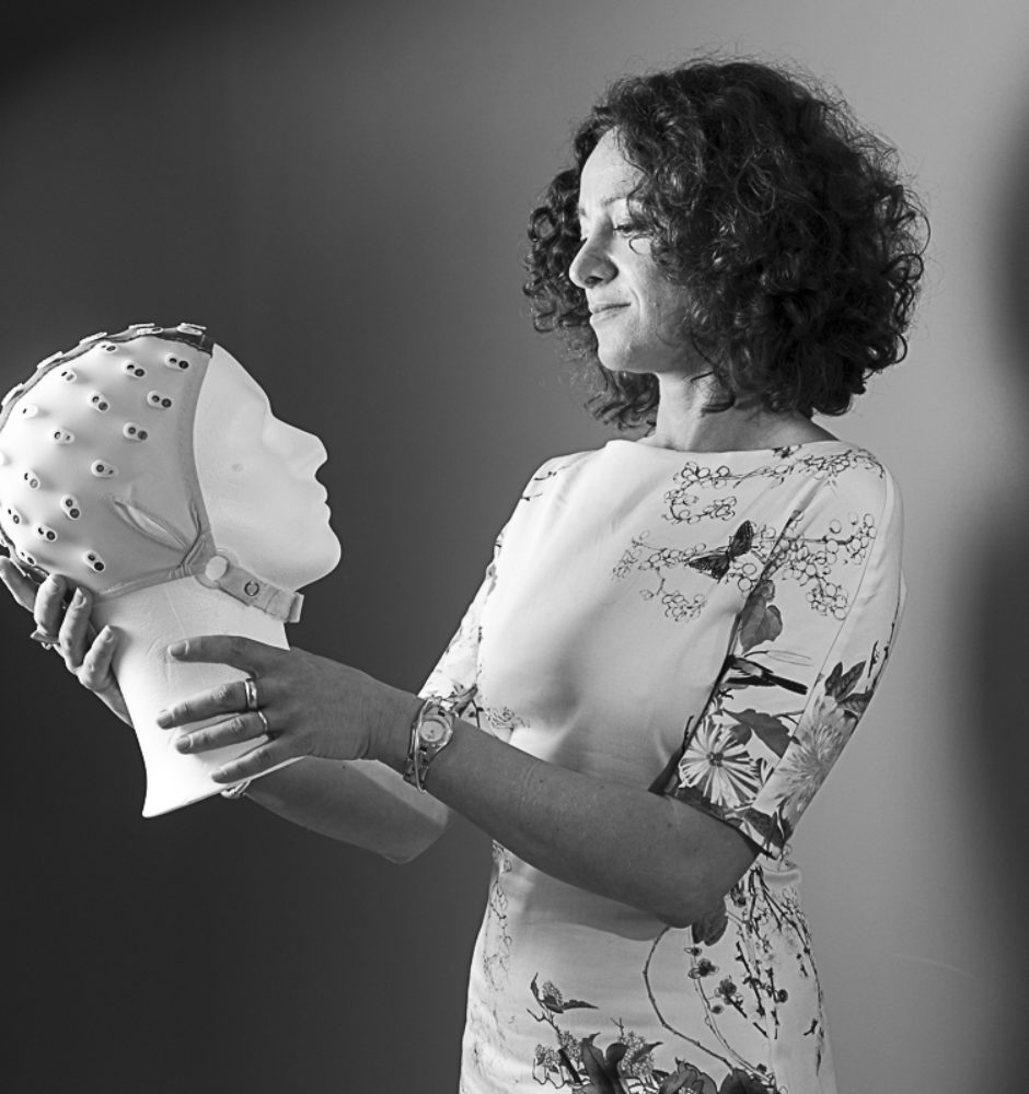
ActiVest
ActiVest - Vestibular functional exploration in humans and non-human primates
State of the art in vestibular activation
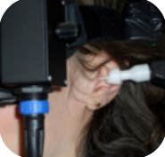
Caloric vestibular stimulation
This method, developed by Robert Bárány, consists of injecting a stream of cold (0.4°C to 20°C) or hot (44°C) water (or air) into the external auditory canal. The temperature difference between the human body and the injected water forms a convection current in the endolymph of the adjacent horizontal semicircular canal. The endolymph flows in opposite directions depending on the temperature used (cold or warm). In a healthy person, this results in a horizontal nystagmus. In the inner ear, activation occurs mainly in the horizontal semicircular canal, with a relatively small contribution from the vertical canal and limited mobilization of the utricle and saccule. This activation propagates to the cerebral cortex, interacting with the neural processes of gravity and inertia signals.
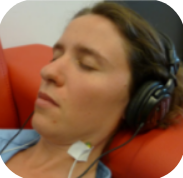
Sound-induced vestibular stimulation
The use of sound-induced vestibular stimulation consists of sending auditory stimuli such as taps or bursts of short sounds into a headset worn by the subject. These short and sudden sounds trigger myogenic potentials
These short and sudden sounds trigger myogenic potentials that can be recorded in the sternocleidomastoid muscles located in the ventral part of the neck. This method mainly activates neural pathways originating from the otolith organs (mainly the saccule, detecting vertical accelerations). This method is used in electrophysiology, but also in functional MRI.
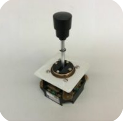
Vestibular stimulation by sled or rotary chair
The rotatory chair is a structure that allows to limit or not the movements of the subject who is sitting in it. Between the device and the head of the subject, a vertical rod allows to measure continuously the position of the head, using a coil. An electrophysiological measurement system can be linked to this same device via a hydraulic device. A screen can be placed in front of the subject to use visual cues or elicit eye movements. It is then possible to record the head movements actively performed by the subject in response to the stimuli. These movements can then be reproduced passively by controlling the chair movements, while blocking voluntary movements.
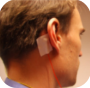
Galvanic vestibular stimulation
Galvanic vestibular stimulation is the method preferentially used by our team. It consists of the application of a weak percutaneous current through an anode and a cathode, placed on the opposite mastoid apophyses (behind the ear). Although low in intensity, this current is sufficient to modulate the level of hyperpolarization of the vestibular neuroepithelia. The excitation rate increases in the vestibular afferents ipsilateral to the cathode, while it decreases on the anode side. This method of stimulation has the advantage of offering different configurations to elicit a specific movement: forward, backward, left or right.
photo: Lopez, Blanke and Mast, “The human vestibular cortex revealed by coordinate-based activation likelihood estimation meta-analysis”, 2012
Team objectives
• Compare the different vestibular activation techniques
• Describe the effects of galvanic stimulation on different functions: postural, oculomotor, perceptual and cerebral
• Describe the mode of action of galvanic stimulation according to the parameters of stimulation: frequency, intensity, duration and shape of the
stimulus
To be able to propose with the support of fundamental data the best stimulation pattern for each of the major functions in which all or part of the vestibular system is involved.
Methods and techniques
Once the most suitable vestibular activation method has been chosen (galvanic vestibular stimulation for our theme), it is important to be able to attest to the subject’s suitable response, both at the cerebral and behavioral levels. Once this proof of effectiveness of the stimulation is obtained, these same methods are used to measure the responses of the subjects during the experimental protocols.
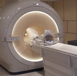
Functional imaging
by magnetic resonance (fMRI)
fMRI is a brain imaging technique that measures the activity of brain areas in vivo. It is based on a simple biological concept: when a brain area is activated, it requires a greater blood flow to cover the metabolic needs generated. This influx will induce a modification of the ratio between oxyhemoglobin and deoxyhemoglobin, leading to the appearance of a magnetic signal measurable in MRI, the BOLD signal. MRI can also be used to measure the connectivity between different brain regions in a subject at rest or more simply to obtain an anatomical image of the brain.
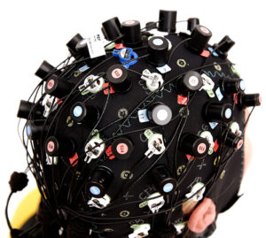
Functional near infrared spectroscopic imaging (fNIRS)
fNIRS is the application of near-infrared spectroscopy to brain imaging. This method allows to measure the BOLD signal, just like fMRI, i.e. to measure the oxygenation of a cerebral area and to deduce its activity level. fNIRS is complementary to fMRI in that it allows the subject to be placed in several postures, unlike fMRI which requires the patient to be lying down. On the other hand, fNIRS works via a cap like EEG and does not allow for a great depth of measurement and will therefore be used more for the analysis of cortical areas.

Psychophysical measurements
Psychophysics can be defined as a measure of the ability to detect a signal, or to differentiate between two stimuli of the same nature if their intensities are very close. They are carried out by providing the subject with a system allowing him to respond (button, joystick, ...) to a stimulation. The psychophysical measurement completes the biological measurements (fMRI and fNIRS) by giving a subjective measurement of the subject's perception. This method is essential in addition to the previous ones to ensure that the subject consciously perceives the stimulus induced during the experiment.
Activest members
Alexandra Séverac Cauquil
CerCo CNRS/UT3
Alexandra Séverac Cauquil, CerCo/UT3 senior lecturer has an international expertise on the vestibular system, visuo-vestibular integration in humans and is a leader in vestibular galvanic stimulation in France. She has previously coordinated projects on vestibular rehabilitation through GVS, visuo-vestibular interactions for posture, post-stroke vision recovery and 3D vision in Parkinson's disease. Her current project within the SV3M team at CerCo focuses on galvanic vestibular stimulation and functional MRI identification of cortical visuo-vestibular integration. She coordinates the project "In-Vest" funded by the ANR-21-CE37-0023-01.
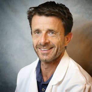
Stéphane Besnard
Univ. de Caen-Normandie
Stéphane Besnard is a professor of physiology at the University and Faculty of Medicine of Caen and a neurophysiologist at the University Hospital of Caen. He is an associate researcher at the LNCS and was a member of the COMETE unit for 12 years. He has a PhD in medicine and neuroscience. His research interests include the vestibular system, multisensory integration and motion sickness, as well as related biotechnology development. His projects in physiology, neuroscience and neurophysiology involve human and mouse models. He was the head of the medical committee for parabolic flights and is currently involved in the DeepTime project. He is also the co-founder of two MEDTECH start-ups.

Jean-Baptiste Durand
CerCo CNRS/UT3
Jean-Baptiste Durand is a CNRS researcher at CerCo since October 2010. After a master's degree in neurophysiology and neuroscience at UT3, and a PhD at CerCo on the cortical processing of three-dimensional visual space in the primary visual area of the vigilant monkey, he completed a post-doctoral fellowship at the Catholic University of Leuven in Belgium before joining CerCo as a research fellow. He currently co-leads the SV3M team and studies the visual system in both human and non-human primates. His research is based on a comparative approach man/ape and relies on complementary techniques: psychophysics, functional imaging (IRMf) and electrophysiology.
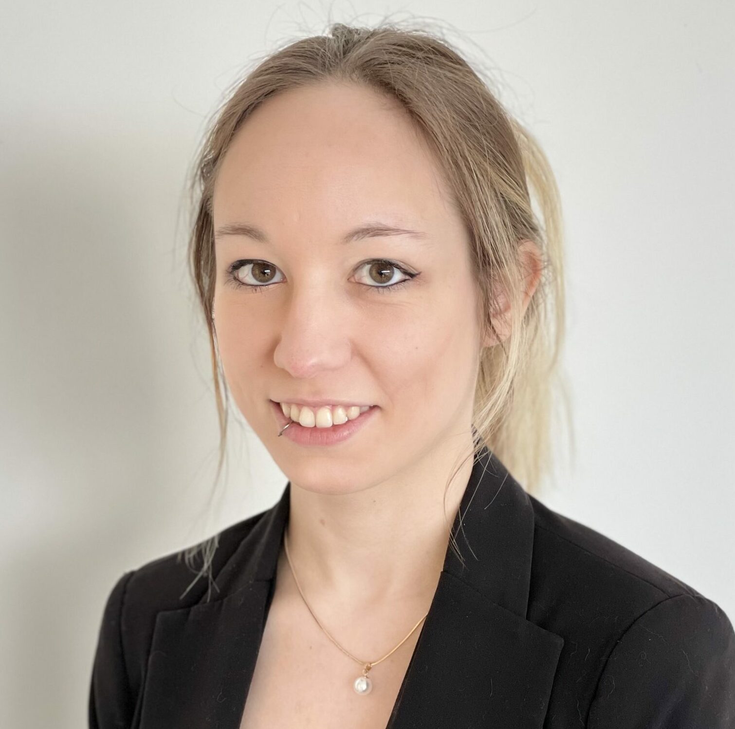
Sarah Marchand
CerCo CNRS/UT3
Sarah Marchand is a PhD student at CerCo since January 2022. After a master's degree in Neurosciences, Behavior, Cognition at UT3, she joined the CerCo and the SV3M team to carry out her thesis under the co-direction of Alexandra Séverac Cauquil and Jean-Baptise Durand. During her first research experiences, she worked with very diverse models (ovine, caprine, murine, canine and feline) on issues related to parasitology, animal cognition and social intelligence. She has a strong interest in comparative cognition and her current project focuses on the integration of visual and vestibular information in a comparative human and non-human primate study using fMRI.
Recent papers
Connectivity of the cingulate sulcus visual area (CSv) in macaque monkeys
V De Castro, AT Smith, AL Beer, C Leguen, Nathalie Vayssière, Yseult Héjja-Brichard, P Audurier, BR Cottereau, JB Durand
Vestibular Modulation of Long-Term Potentiation and NMDA Receptor Expression in the Hippocampus
Paul F. Smith, Bruno Truchet, Franck A. Chaillan, Yiwen Zheng, Stephane Besnard
Vestibular stimulation by 2G hypergravity modifies resynchronization in temperature rhythm in rats
Tristan Martin, Tristan Bonargent, Stéphane Besnard, Gaëlle Quarck, Benoit Mauvieux, Eric Pigeon, Pierre Denise & Damien Davenne
Dynamics of the straight-ahead preference in human visual cortex
Olena V Bogdanova, Volodymyr B Bogdanov, Jean-Baptiste Durand, Yves Trotter, Benoit R Cottereau
Antero-Posterior vs. Lateral Vestibular Input Processing in Human Visual Cortex
V6 is active during antero-posterior but not in lateral galvanic vestibular stimulation
Felipe Aedo-Jury, Simona Celebrini, Benoit Cottereau, Maxime Rosito, Alexandra Séverac-Cauquil
Ongoing projects
Find in this section the projects carried out in parallel by the different members of the ActiVest team.
In-Vest
In-Vest is a project coordinated by Alexandra Séverac Cauquil. It is a collaboration between CerCo (CNRS/UT3, Toulouse), CLLE (CNRS/UT2J, Toulouse) and VERTEX (Univ. Caen-Normandie). The objective is to better define the functioning of the vestibular system, by developing a research and rehabilitation tool to stimulate the vestibular system, which during this project, will be used to understand the role of factors such as aging or posture on the activation of the vestibular system.
Deep Time
Stéphane Besnard coordinates the research program of the "Deep time" expedition.
One year after the first confinement, 15 women and men between 27 and 50 years old live for 40 days in a cave in Ariège without any notion of time. They are equipped with sensors allowing a dozen scientists to follow them from the surface. The objective is to analyze the individual behavior and the emotional and cognitive capacities, as well as the physiology.

May 9-11, 2022
XXXI Bárány Society Meeting
The congress will focus on major breakthroughs and advances in vestibular medicine and will provide an innovative and comprehensive overview of the latest research developments, primarily in the areas of molecular biology, neurophysiology and imaging diagnostics, medical and surgical treatment, and rehabilitation. Some of these objectives will be achieved at the Vestibular Science Satellite Meeting in the city of Granada to be held on September 4-5, 2020, prior to the main meeting.
